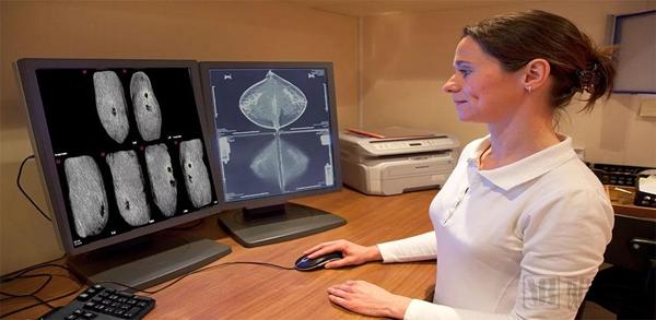
Breast cancer is one of the most important diseases that cause death in women today. The incidence of breast cancer in China is also increasing year by year, and it is now the highest in female malignant tumors. Studies have shown that breast cancer is a disease that affects the whole body and has a high mortality rate. Humans are not powerless. If they can find the disease early in the cancer, after effective treatment, women can not only avoid pain, but also prevent the spread of cancer cells.
Nowadays, a large number of patients are diagnosed by performing breast DR photography (also known as mammography). Usually, these results are reviewed by doctors one by one, which leads to a significant increase in the workload of the reading doctors, and it is more difficult to find the lesion area.
Computer-aided diagnosis (CAD) is a new technology applied to the field of imaging technology with the development of computer technology and digital medical in recent years. It uses professional computer algorithms to analyze images, find and detect lesion characteristics, and help radiologists improve the detection rate of lesions.
As a leader in the implementation of ultrasound CAD screening for dense breast tissue, Qview Medical has developed a CAD system for 3D comprehensive breast ultrasound that has been upgraded and upgraded on the original CAD imaging technology.
According to the arterial network, the company's product QVCAD is based on a deep learning algorithm that combines innovative C-Thru technology with ABUS to reduce image read time by 33% while maintaining diagnostic accuracy.
Fully known as Mammography Automated Volume Imaging, ABUS provides one-button standardized workflow and image reading, including standardization operations, standardized imaging, standardized reporting, and automatic generation of standardized, full-volume ultrasound images. The study found that breast cancer detection was increased by 35.7% compared with mammography alone.
Founded in 2006, QView Medical has extensive application experience in the field of artificial intelligence (AI). In November 2016, the team developed the first PMA-approved CAD system for mammography and lung CT. PMA, also known as pre-market approval, is a scientific regulatory assessment of the safety and effectiveness of medical devices by the FDA (US Food and Drug Administration).
QView Medical has conducted in-depth research on the development of QVCAD algorithms for 3D ABUS while collecting cancer cases from more than 1 million ABUS 3D images worldwide. The company said that the solution provided by QVCAD Invenia ABUS has the potential to be the preferred screening method for women with dense breasts.
In December 2017, QView Medical announced that FDA approval for QVCAD and GE Invenia ABUS systems for women's dense breast testing.
In March 2015, QView Medical received $4.8 million in risk round financing, which was not disclosed by the investors.
Years of entrepreneurial experience to promote breast imaging development
Bob Wang, CEO and Chairman of QView Medical, has established several companies that have transformed medical imaging and improved breast care over the past 30 years. His outstanding vision and action in the imaging industry has driven the development and advancement of modern mammography.
Wang said that when he first came into contact with medical imaging, the high-radiation exposure of mammography was considered harmful to the human body and could not be screened for breast. Under Wang's in-depth study, his high-resolution, low-dose rare-earth X-ray photography technology was successfully sold to 3M and licensed by Kodak.
Wang reduced the dose of mammography by 95% and evolved into the breast imaging technology we use today. According to the arterial network, this rare earth technology, which can reduce the X-ray dose, can be used for all X-ray photography procedures worldwide, worth billions of dollars.
From 1993 to 2006, Wang founded R2 Technology and served as the company's CEO and chairman. The R2 technology focuses on the impact of CAD systems on early detection of breast cancer and is committed to commercialization. At the same time, this is the first example of the world to introduce CAD into mammography and is widely used in various countries. In July 2006, R2 Technology was acquired by Hologic.
Wang did not stop further learning and inquiry into medical imaging. He observed that mammography did not reflect the key features of early breast cancer in the face of breast dense cells. On the contrary, ultrasound examination seems to be more effective.
In January 1997, Wang established U-Systems as the company's CEO and chairman. U-Systems primarily designs and develops breast screening system products and was acquired by GE Healthcare in November 2012. Then, Wang developed the operation somo. The important technology of v. Somo. v is an automated breast ultrasound system that has also become the first FDA-approved ultrasound breast examination device.
In addition, Wang has received degrees from the Massachusetts Institute of Technology and Rensselaer Polytechnic Institute. In January 2011, he founded EndoSee Corporation, a company selling medical equipment , by The Cooper Companies Inc. Acquired in California, USA for $44 million. Cooper plans to bring high-quality products into the office/surgical strategy of obstetrics and gynecology practice (OB / GYN) while leveraging EndoSee's infrastructure.
Wang established QView Medical and has established W&Wsens Devices and UroViu Corporation in November 2014 and March 2016 respectively. UroViu's product, the disposable diagnostic cystoscopy system (Uro-V), has been licensed and patented by the FDA under 510(k).
Ron Ho, another key member of QView Medical, is a member of the board of directors of the company. Not long ago, Ron Ho became the new CEO of U-Systems. Prior to joining U-Systems, Ron Ho led laser scanning company Metron Systems to provide 3D digital precision technology for complex parts in the medical, aerospace and aerospace industries. At the same time, he has 16 years of experience in the ultrasonic group of Siemens Medical Solutions, responsible for product development and product output of the company's ultrasonic transducers.
Currently, Ron Ho is a director of several medical device companies and holds a bachelor's degree in mechanical engineering (BSME) and a master's degree in mechanical engineering (MSME) from the University of Washington. According to the arterial network (WeChat: vcbeat), he has obtained a number of related patents in the field of ultrasound.
What is the principle of the QVCAD system?
As the first CAD system to develop 3D automatic breast ultrasound, QView Medical is mainly used to screen dense breast tissue for precise diagnosis and treatment. Its product QVCAD system helps radiologists greatly improve breast screening efficiency. The QVCAD system is especially aimed at patients who have a negative result in mammography and who have used the 3D breast ultrasound ABUS system under the influence of dense breast tissue. The multi-modal combination of mammography and breast ultrasound provides a solution for women with dense breasts.
In addition, QView Medical recognizes the value of the ABUS system. They believe that the efficiency of machine reading must be improved while maintaining diagnostic precision. With the company's deep learning in the AI ​​field, they developed new algorithms on the original ABUS system and rigorously tested them to improve machine reading for the ultimate purpose.
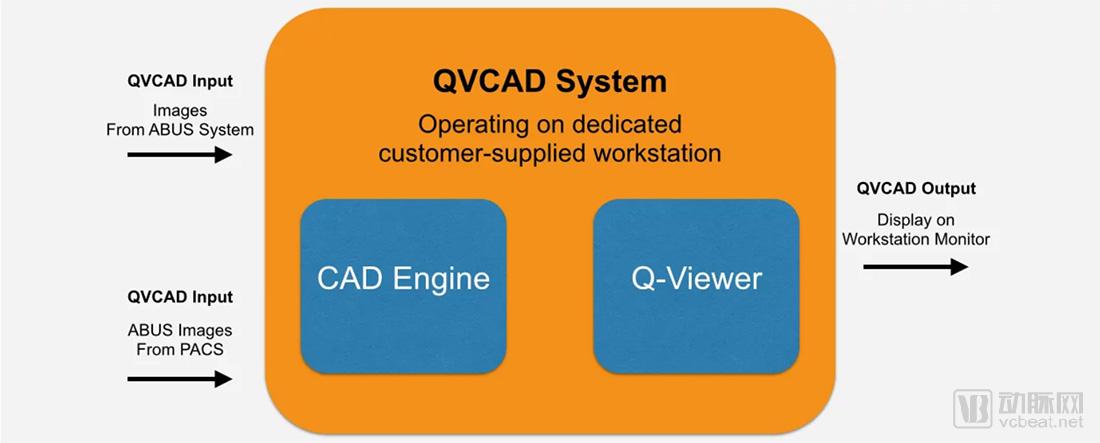
QVCAD is a software system consisting of two subsystems:
1. The CAD subsystem contains complex image processing methods;
2. The Viewer subsystem recombines the ABUS image with the output of the QView CAD engine to display the image on the display for medical personnel to read.
The QVCAD system receives input from ABUS images and ABUS images from PACs systems via the standard DICOM format. The native image of the ABUS system can display the output image on the Q-Viewer as it passes through the QVCAD CAD engine.
The QVCAD CAD engine uses a variety of image pattern recognition processes and uses artificial neural networks to detect suspicious lesions in the breast. Its main purpose is to distinguish potential breast lesions from normal breast tissue.
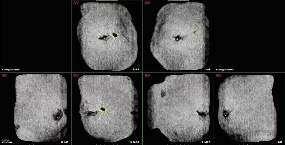
The CAD engine output of the QVCAD system is mainly presented in the following two ways:
1. Presented in a coronal image format. As shown in the navigation image of QVCAD below, CAD has detected possible abnormalities in the presence of the breast. The key parts of the CAD mark are represented by the green circle in the figure, and the content is directly displayed by the CAD image navigator. The CAD mark emphasizes potential malignant lesions.
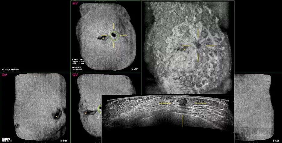
2. Cursor hover mode allows the user to quickly view the localized area of ​​the view. The method is to display the original image of ABUS Coronal and Transverse corresponding to the CAD navigation image, and observe the point at which the cursor hovers. The hover mode is activated when the cursor is over any area of ​​the CAD navigation image.
QView Medical vs. competitors
1. Comparison of QView Medical and iCAD
Founded in 1984, iCAD is headquartered in Nashua, New Hampshire, USA, and is dedicated to providing advanced image analysis, workflow solutions, and radiation therapy for early identification and treatment of cancer. The company operates primarily through the cancer detection and cancer treatment departments. Cancer testing includes image analysis and solutions to support clinical decision making for mammography, mammary tomosynthesis and computed tomography imaging. Cancer treatment mainly provides platform technology without isotope cancer treatment.
Compared with Qview Medical, iCAD is established early, has many products and covers a wide range of diseases, providing patients with a full range of services for cancer detection and treatment. iCAD's PowerLook Breast Health Solutions offers 2D and 3D mammography combined with AI technology to meet patient needs.
2. Comparison of QView Medical and Volpara
Volpara is committed to providing digital medical solutions for the early detection of breast cancer, which can better analyze the density of female breasts and analyze and screen them based on objective measurements of breast density, compression and radiation dose. Volpara Solutions has developed several software. Among them, VolparaDensity can provide clinicians with patient-specific X-ray radiation dose through mammography diagnosis, and convert mammography into volumetric data for clinical decision making. Volpara's technology has been applied in more than 30 countries and its coverage will continue to expand.
Compared with Qview Medical, Volpara mainly measures and analyzes women's breast density through the transformation of images and data. Qview Medical provides solutions for dense breast women for the upgrade and replacement of image and ultrasound technology.
Suyzeko Photobiomodulation Therapy Helmet 810nm Infrared Helmet PBM Light Therapy Machine
infrared red light helmet 810nm Photobiomodulation benefits and photobiomodulation therapy near me
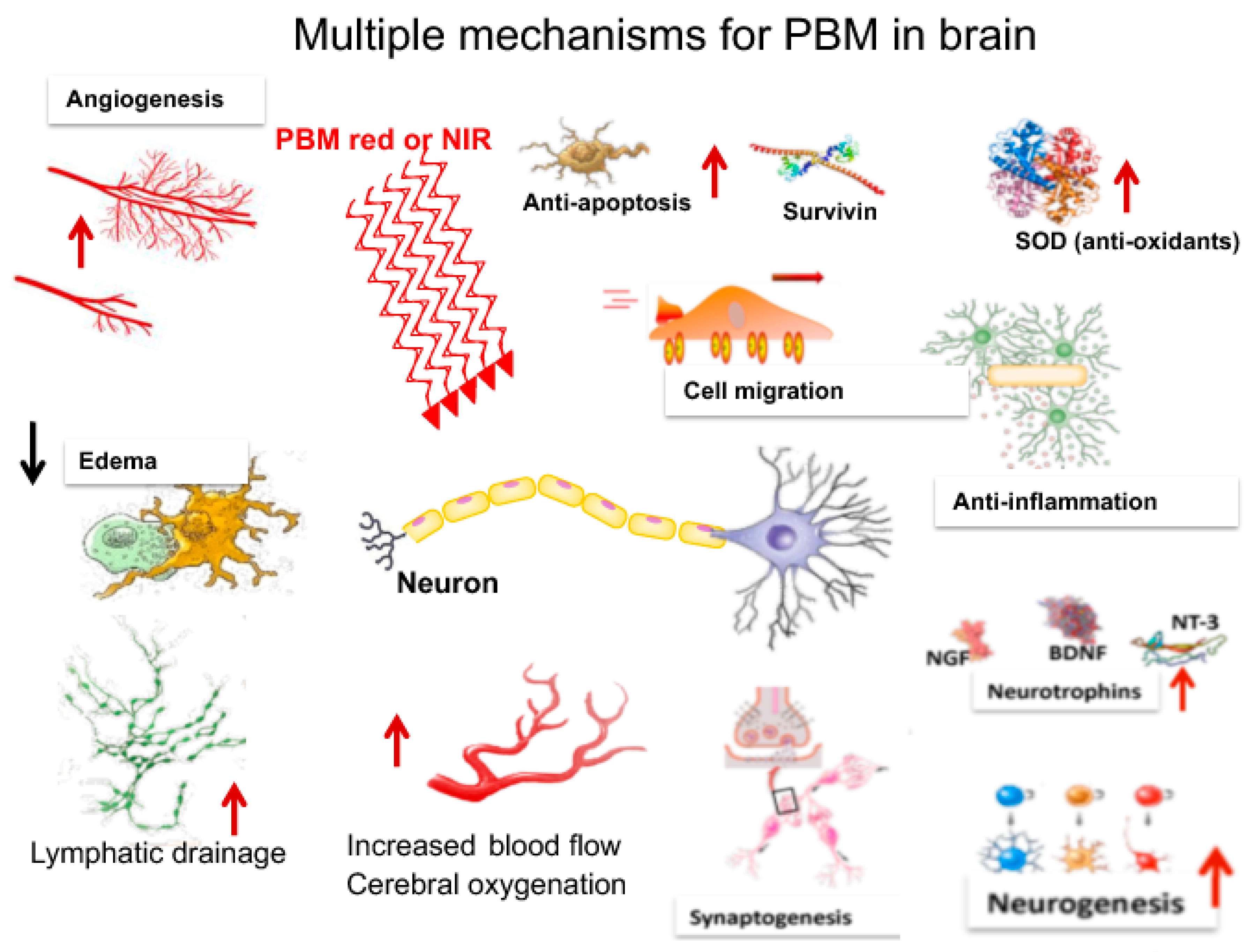
Photobiomodulation (PBM) describes the use of red or near-infrared light to stimulate, heal, regenerate, and protect tissue that has either been injured, is degenerating, or else is at risk of dying. One of the organ systems of the human body that is most necessary to life, and whose optimum functioning is most worried about by humankind in general, is the brain.
PBM light therapy, also known as photobiomodulation therapy, has been shown to have several benefits for brain disorders. This therapy involves using low-level light therapy to stimulate the cells in the brain and promote healing and regeneration. Here are some of the benefits of PBM light therapy for brain disorders:
1. Neuroprotection: PBM light therapy has been found to have neuroprotective effects, meaning it can protect the brain cells from damage and degeneration. It can help reduce inflammation and oxidative stress in the brain, which are common factors in many brain disorders.
2. Improved cognitive function: PBM light therapy has been shown to enhance cognitive function, including memory, attention, and problem-solving skills. It can stimulate the production of brain-derived neurotrophic factor (BDNF), a protein that promotes the growth and survival of brain cells.
3. Reduced symptoms of depression and anxiety: PBM light therapy has been found to have antidepressant and anxiolytic effects. It can increase the production of serotonin, a neurotransmitter that regulates mood, and reduce the activity of the amygdala, a brain region involved in fear and anxiety.
4. Enhanced neuroplasticity: PBM light therapy can promote neuroplasticity, which is the brain's ability to reorganize and form new connections. This can be beneficial for individuals with brain disorders as it can help improve their ability to learn, adapt, and recover from injuries.
5. Accelerated healing after brain injuries: PBM light therapy has been shown to accelerate the healing process after brain injuries, such as traumatic brain injury or stroke. It can improve blood flow to the injured area, reduce inflammation, and stimulate the repair and regeneration of damaged brain cells.
photobiomodulation, pbm light therapy,photobiomodulation therapy machine,Infrared Helmet,810nm 1070nm pbm helmet, rtms helmet
Shenzhen Guangyang Zhongkang Technology Co., Ltd. , https://www.nirlighttherapy.com